
Sonography Treatment
Introduction
Our Sonography Treatment
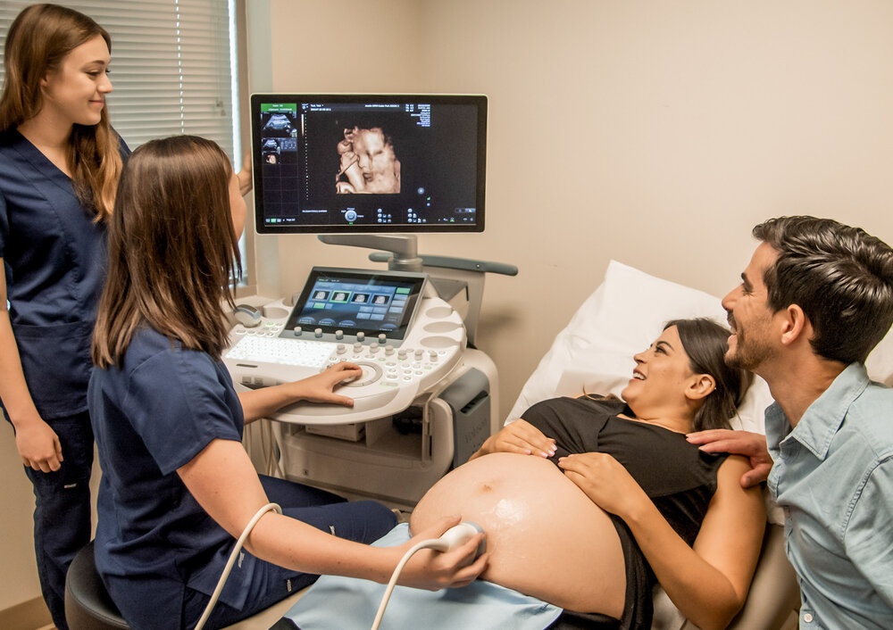
Madhuram Women's & General Hospital using a handheld device called a transducer to transmit high-frequency sound waves into the body. The sound waves bounce off of internal structures and are detected by the transducer, which then sends the information to a computer that converts it into real-time images on a monitor.
Sonography is commonly used to evaluate the health and function of various organs and structures in the body, including the heart, liver, kidneys, uterus, ovaries, and blood vessels. It is a non-invasive, painless, and radiation-free diagnostic tool that is widely used in both diagnostic and therapeutic medicine.
Unlike X-rays or CT scans, which use ionizing radiation to produce images, sonography uses sound waves, which are safe and do not pose any risk of radiation exposure.
Read More
Sonography produces real-time images, which means that doctors and sonographers can see the results immediately and make any necessary adjustments or interventions during the procedure.
Read More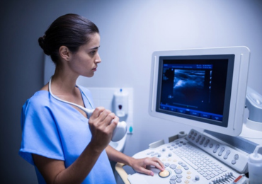
Sonography can be used to evaluate many different parts of the body, including organs, blood vessels, and soft tissues. It can also be used to guide certain procedures, such as needle biopsies.
Read More
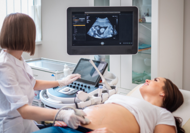
Sonography is a safe diagnostic tool for pregnant women, as it does not use any ionizing radiation and does not pose any risk to the developing fetus.
Read More